Book Appoinment
Laser Varicose Veins Training Program in India
What is EVLA?
EVLA is a method of treating varicose veins using carefully controlled laser to ablate the vein. The veins usually targeted are in the main trunks of the legs:
The veins usually targeted are in the main trunks of the legs:
- Great Saphenous Vein (GSV)
- Small Saphenous Vein (SSV)
- Minimal recurrence
- Tributary Veins such as Anterior Accessory Saphenous Vein (AASV)
Why Laser for Endo Venous Laser Ablation (EVLA)?
Varicose vein is probably the most common lifestyle ailment affecting both men and women at any age. Most patients are known to avoid surgical treatment because it is painful, time-consuming, and worst of all, and creates a scar. Now the patients are well-informed about the use of the laser to treat their conditions and almost demand to use the same for their case. This is not a very surprising situation because it means that your patients are eager to lead a better life, free from pain and discomfort.
If you really want to help more and more patients step towards a better life, start saying “Yes” to Laser surgery. Participate in this unique program designed by Photoniccs for ambitious practitioners like you.
Procedures You Will Learn in EVLA Training
The EVLA laser procedure is carried out by inserting the laser fibre into the affected varicose vein (endo venous means inside the vein). The detailed procedure is as follows:
- Apply a local anaesthetic over the affected area and insert a needle in the area.
- Pass a wire through the needle up the vein.
- Remove the needle and pass a catheter (thin plastic tubing) over the wire into the saphenous vein
- Pass a laser radial fibre up the catheter in such a way that its tip reached the point that needs to be heated the most (usually the groin crease).
- Inject enough local anaesthetic solution into the vein through multiple needle pricks or by Tumescent anaesthesia.
- Fire up the laser and pull the radial fibre down centimetre by centimetre in 20 to 30 minutes.
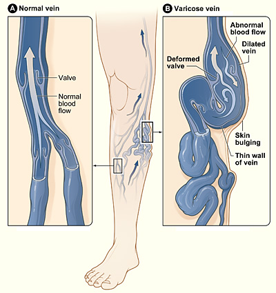
- Heat the veins through by laser fibre passed through the catheter causing homogenous destruction of the vein’s walls by shrinking it and sealing it off. As a result, there is no more blood flow in these veins that may result in swelling. The surrounding healthy veins are free of the varicose veins and therefore able to resume with the healthy blood flow.
- Remove the laser and the catheter and cover the needle puncture wound with a small dressing.
This procedure takes 20 to 30 minutes per leg. The smaller veins may need to undergo sclerotherapy

Our Mentor
Dr. Maunil A. Bhuta
MD | DNB | FENI | MBBS
Interventional Radiologist
General Surgeon & Laparoscopic Surgeon
Dr. Maunil Ajay Bhuta is a Neuro and Vascular Interventional Radiologist. He was awarded the prestigious fellowship in Endovascular Neuro-Interventions – Changhai Hospital, Shanghai.
He has 7 years of experience in CT and USG guided Interventions and 4 years of experience in complex neuro and Vascular interventions. He was an Assistant Professor in the Dept. Neuro of Vascular Interventional Radiologist at Sion Hospital.
Achievements
1. Done more than 1000+ Cases of EVLA for varicose veins
2. 2nd Rank in the MD University Examination (2015)
PUBLICATIONS, PAPERS & POSTERS
Imaging of Pancreatico-Biliary Tumors: CT & MRI (International Journal of Scientific Research–2017)
- Role of uterine artery embolization in the management of menorrhagia (Indian Journal of Basic and Applied Medical Research–2016)
- Study of role of pre-operative embolization of Juvenile Nasopharyngeal Angiofibroma (JNA) tumours (Indian Journal of Basic and Applied Medical Research–2016)
- Role of Neuro-ultra sound in evaluation of intracranial pathologies in preterm neonates and infants with abnormal neurological presentation. (International Journal of Science and Research–2016)
- Role of magnetic resonance venography in the assessment on intracranial venous thrombosis (Indian Journal of Basic and Applied Medical Research – 2016)
- MRI Imaging In Perianal Fistulas: A comparative Study of 25 Patients (World Journal of Pharmaceutical and Medical Research-2016)
- A pictorial essay– Neuro- radiological Manifestations of Neurofibromatosis type II (Indian Journal of Basic and Applied Medical Research – 2016)
- Case of Double Right Coronary Artery: Diagnosed on Computed Tomography Coronary Angiography (Heart India–2015)
- Vision Loss due to Central Retinal Artery Occlusion Following Embolization in a Case of a Giant Juvenile Nasopharyngeal Angiofibroma (Journal of Craniofacial Surgery–2015)
- Vision Loss due to Central Retinal Artery Occlusion Following Embolization in a Case of a Giant Juvenile Nasopharyngeal Angiofibroma (Journal of Craniofacial Surgery–2015)